
A bone recovered on Ireland’s coast became a perfect test case for non-destructive analysis. Using industrial X-ray CT (diondo d5) and SEM/EDX at SEAM, we visualised the bone’s internal architecture and identified its elemental composition—without cutting or damage. Here’s how high-resolution CT and materials analysis can reveal hidden structure, history and environment in archaeological and industrial samples alike.
Revealing Secrets with Industrial X-ray CT and Material Analysis
What happens when cutting-edge technology meets ancient mystery?
A recent archaeological test on a bone found along the Irish coast shows how industrial X-ray CT imaging and material analysis can uncover the untold story locked inside a seemingly ordinary object.
Thanks to advanced tools like the diondo d5 CT system and SEAM (Scanning Electron Analysis of Materials), we were able to non-destructively visualize the internal architecture of the bone and identify its elemental composition—all without damaging this precious find.
High-Resolution X-ray Imaging with diondo d5
To explore the internal structure of the bone, we used the diondo d5 industrial CT system, equipped with a versatile range of X-ray tubes and a high-performance detector:
- A 240 kV microfocus X-ray tube, used in this experiment, provided ultra-fine detail for capturing the bone’s internal features
- 450 kV mesofocus and minifocus tubes offer greater penetration power, ideal for scanning denser or larger materials
- A 3000-pixel detector with a 139-micron pixel pitch enables high-resolution imaging across a wide range of sample sizes
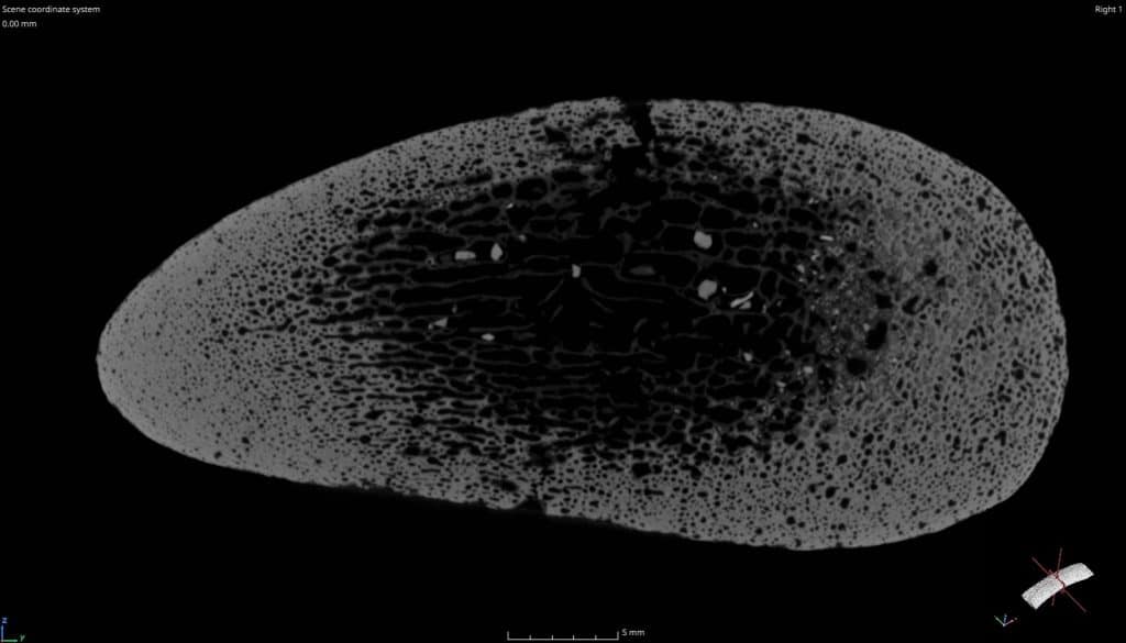
In this particular test, we scanned a 16 cm section of a larger 40 cm-long bone. Operating at 4× magnification, the system achieved a voxel resolution of just 35 microns, delivering a remarkably detailed view of the bone’s internal complexity.
What the CT Revealed
The 3D reconstructions uncovered:
- A dense cortical layer—the outer shell of the bone
- An internal network of trabeculae (thin, rod-like structures) and marrow cavities, characteristic of cancellous (spongy) bone
- Bright white inclusions scattered throughout the internal structure
These bright spots indicated the presence of dense foreign materials—likely shell fragments, or marine minerals that infiltrated the bone over time. This aligns with the bone’s origin from a marine environment, suggesting long-term exposure to the sea and sediment.
See CT Scan Video Here
SEM/EDX Findings: Hydroxyapatite, Silica & Marine Trace Elements
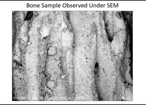
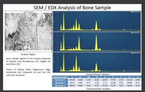
To dig deeper (without physically digging), we used Scanning Electron Microscopy (SEM) combined with Energy-Dispersive X-ray Spectroscopy (EDX). This dual technique allows for pinpoint identification of elemental composition at micro-scale resolution.
Key Findings:
- High levels of Calcium (Ca), Phosphorus (P), and Oxygen (O) – the foundational elements of hydroxyapatite, the mineral that makes up natural bone.
- Silicon (Si) – possibly absorbed sand or silica from the marine environment.
- Trace elements such as Sodium (Na), Magnesium (Mg), Aluminum (Al), Potassium (K), and Iron (Fe) – likely introduced by seawater or sediment over time.
Together, these findings confirm the authenticity of the bone and reveal how it has chemically interacted with its marine surroundings, absorbing minerals and evolving over time.
What SEAM Can We Test – Without Cutting or Damaging the Sample?
One of the most exciting aspects of this work is that all of it was done non-destructively. That’s the true power of industrial CT imaging:
✅ X-ray or CT Scanning – See inside any object without slicing it open
✅ Material Analysis (SEM/EDX) – Determine what it’s made of on an elemental level
Final Thoughts: A Journey from Beach to Lab
From examining industrial parts and medical prototypes to scanning an ancient bone, this project was a refreshing dive into the natural sciences.
The bone—possibly part of a vertebral column or horn core—has yet to be definitively identified, but the imaging and analysis have already told part of its story. A story shaped by biology, time, and the relentless forces of the sea.
So next time you walk along the beach and spot something unusual, remember—what looks like a weathered fragment may just be a doorway into the past, waiting to be revealed with the right technology.
Want to Explore What’s Inside Your Sample?
Whether it’s archaeological, or industrial, our non-destructive testing solutions give you deep insights without damaging what matters.
➡️ Discover what’s hidden inside.
➡️ Preserve while you analyze.
➡️ Contact us to learn more about industrial CT and material analysis services.
This piece was written by Agnieszka Furman – Technology Leader & Business Development X-ray Micro Computed Tomography . If you would like to learn more about our CT or Non destructive testing capability at SEAM please contact us.